Expert Doctor Specializing in Treating Melasma and Dark Spots in Bangkok-BAC Clinic


Darks spots, moles, or hyperpigmentation are conditions where certain areas of the skin become darker than usual or develop excess pigmentation in those areas, resulting in an uneven complexion with dark patches that are difficult to conceal. While not necessarily severe, these conditions can significantly impact confidence and self-image, often requiring constant cosmetic coverage. Both internal hormonal factors and external environmental conditions contribute to the onset of these conditions. Therefore, understanding the causes and treatment options can help individuals make better decisions in self-care and prevention.
What are melasma? Melasma are a common dermatological condition caused by the overactivity of melanocytes, the cells responsible for producing pigment in the skin. This leads to an increase in localized pigment production (hyperpigmentation), resulting in darker patches, especially prominent on the face and sometimes on the neck, chest, and arms. Freckles are most prevalent among individuals with Fitzpatrick skin types III-IV, commonly found in Asian populations. They appear as light to dark brown spots or patches and are more frequently observed in women than in men, with a ratio of 9:1.
The exact cause of melasma remains unclear, but it is believed to be associated with the overactivity of melanocytes beneath the skin’s surface. Freckles occur due to an increased melanin production, with melanocytes in affected areas having longer dendrites and a higher concentration of melanosomes in stages III and IV, both in epidermal and dermal melanocytes. Factors that increase the likelihood of freckles include Asian ethnicity, particularly Fitzpatrick skin types III-IV, female gender, sun exposure, hormonal changes such as during pregnancy or with contraceptive use, genetics, thyroid disorders, and low levels of testosterone.
In summary, understanding the factors contributing to the development of freckles can help individuals take proactive measures to manage and prevent these conditions, thereby improving their self-care and confidence.
Periorbital hyperpigmentation, also known as dark circles under the eyes, occurs due to the thinness of the periorbital skin, leading to the visibility of blood vessels, hollows, and shadows. Accumulation of melanin in the dermis, post-inflammatory hyperpigmentation (PIH) resulting from conditions like allergic contact dermatitis, etc., can contribute to its formation. Treatment options for periorbital hyperpigmentation include topical medications such as hydroquinone, kojic acid, azelaic acid, etc., as well as various procedures like chemical peels, radiofrequency, fractional laser, picosecond laser, filler injections, fat injections, etc.
Freckles, also known as ephelides, are small, flat, brown spots that appear on the skin due to increased melanin production. They are commonly found on sun-exposed areas of the skin and are usually harmless. Freckles may be treated with topical creams containing ingredients such as hydroquinone, retinoids, or azelaic acid, or through procedures like chemical peels, laser therapy, or cryotherapy, depending on the individual’s preference and severity of the freckles.
Ephelides, freckles

(Ephelides (Freckles) — Dermatology Advisor, n.d.)
These are light brown patches with distinct borders, typically larger than 5 millimeters in size, commonly found in areas exposed to sunlight such as the face, back of the hands, and arms. This condition is more prevalent in individuals with fair skin compared to those with darker skin tones. Prolonged sun exposure tends to darken these spots and may contribute to their increased occurrence.
Solar lentigo, actinic lentigo, sun spot, senile lentigo

(Solar Lentigines — Dermoscopedia, n.d.)
Due to sun exposure, freckles are commonly found on sun-exposed areas such as the face, upper chest, back of the hands, and arms, often seen under shaded areas of clothing exposed to sunlight. Initially, freckles tend to be smaller than 5 millimeters in size, with a light brown color that may darken over time. Freckles usually increase in number gradually and may enlarge in size, with small spots merging to form larger ones. Treatment can involve targeted laser therapy aimed at specific pigmented spots, causing them to peel off gradually within about 7 days. Typically, 1–5 treatment sessions are needed, with freckles often responding well to laser treatment.
Hori’s nevus

(Tian, 2015)
Hori’s nevus typically appear as dark brown or grayish round spots. They are often found in clusters on both sides of the cheeks and both temples, commonly starting around the ages of 20–30. Genetics play a role, with a higher prevalence in females at a ratio of 21:1 compared to males. They resemble freckles but tend to have a more grayish hue, hence the name “Hori’s nevus”
These spots do not fade away on their own and often darken over time. Treatment typically involves targeted laser therapy to remove both epidermal (top layer) and dermal (deeper layer) pigmentation. Picosecond laser is effective, with a minimum of 5 sessions required due to the deep-seated nature of lentigines. Topical treatments or superficial laser therapy targeting only the epidermal layer are ineffective in eliminating lentigines since the melanin deposits are deep within the dermis or inner layers of the skin. Laser treatment may cause transient hyperpigmentation, which typically fades after using whitening agents.
Red and Dark spots from acne

Post-inflammatory erythema (PIE) and post-inflammatory hyperpigmentation (PIH) are residual marks that occur after the inflammation of acne. PIE appears as red marks resulting from inflammation of the underlying tissue combined with the expansion of blood vessels. On the other hand, if inflammation persists over a prolonged period, it stimulates melanocytes to produce excess melanin, resulting in post-inflammatory hyperpigmentation or dark spots after the acne heals. Therefore, gradual and consistent care of inflamed acne can help reduce the severity of acne marks.
Melanocytes are cells that produce melanin, the natural pigment that determines skin color, hair color, eye color, and post-acne dark spots. These dark spots occur due to excessive melanin production or the release of excessive melanin produced by melanocytes, making the dark spots darker than other areas. Thus, individuals with fair skin tend to have a faster fading of dark spots and red marks compared to those with darker skin, as dark spots may linger longer in the latter.
Basic treatment for dark spots usually involves skincare products containing ingredients such as niacinamide, vitamin C, retinol, etc. However, noticeable improvement may take some time and patience.
Currently, the most effective and fastest method for removing dark spots and post-acne marks is Picosecond Laser, the latest innovation in laser technology. It directly affects and destroys melanin granules within seconds, using energy at 1/1,000th of the nanometer level of the traditional Q-switched laser system. Treatment typically requires about 3–5 sessions, depending on the severity of the individual’s condition.
Treatment options for mild to moderate hyperpigmentation, such as post-inflammatory erythema (PIE) and post-inflammatory hyperpigmentation (PIH), include:
Skin exfoliation:
- Chemical peels: Chemicals are applied to the skin to exfoliate the top layer, revealing new, healthier skin underneath.
- Microdermabrasion: A minimally invasive procedure that uses a diamond-tipped wand to gently exfoliate the skin, promoting cell turnover.
Stimulating skin cell turnover in the dermis:
- Vitamin A acid: Also known as retinoids, these compounds promote skin cell turnover and help fade pigmentation.
- Glycolic acid: A type of alpha hydroxy acid (AHA) that exfoliates the skin and encourages the growth of new, evenly pigmented skin cells.
- Salicylic acid: A beta hydroxy acid (BHA) that penetrates deep into the pores to unclog them and improve skin texture
Inhibiting melanin synthesis:
- Hydroquinone: A popular skin-lightening agent that inhibits melanin production.
- Arbutin, kojic acid, licorice extract, tranexamic acid: Other skin-lightening agents that may be effective but are less commonly used due to lower efficacy and higher cost.
Reducing melanin transfer from melanocytes to keratinocytes:
- Laser treatments: Q-switched laser, fractional laser, and picosecond laser are popular options that can target melanin deposits within the skin.
It’s important to note that prolonged use of hydroquinone can lead to rebound hyperpigmentation, where the skin becomes darker than before. Therefore, treatment goals should focus on reducing existing pigmentation, inhibiting the formation of new pigmentation, and preventing future pigmentation by avoiding UV radiation exposure.
While there is no definitive cure for hyperpigmentation, laser treatments have become increasingly popular due to their ability to target melanin deposits effectively. These lasers should ideally have:
- Short pulse width in the nanosecond or picosecond range.
- Adjustable energy levels.
- Wavelengths in the visible to near-infrared range (approximately 1,100 nm).
Studies on ablative laser resurfacing for treating hyperpigmentation have shown that it can cause discomfort, prolonged downtime, and potential uneven skin tone afterwards. Severe side effects such as scarring may also occur. Therefore, to ensure effective treatment, laser penetration into the dermis is necessary to target the root cause of pigmentation. Skin resurfacing with lasers that induce skin peeling (e.g., CO2 or Erbium YAG laser) may provide better results, although they come with longer downtime and a higher risk of side effects.
In the treatment of pigmentation abnormalities, lasers based on the principles of Selective Photothermolysis, which generate heat at the targeted area, have been explored. However, outcomes using lasers such as 532 nm frequency-doubled Q-switched Nd: YAG, 694 nm Q-Switched ruby, or 755 nm Q-switched alexandrite have been unsatisfactory. Similarly, the use of Intense Pulsed Light (IPL), which has a broad spectrum ranging from 515 to 1,200 nm, has been widely employed. Unlike lasers, IPL does not require specific targeting. Its light waves can penetrate both the superficial and deep layers of the skin, with a pulse duration in the millisecond range, allowing for effective heat dispersion and reduced risk of side effects from excessive heat. However, IPL treatment for pigmentation abnormalities should be combined with topical therapy, such as hydroquinone creams, especially for treating superficial and mixed pigmentation types, to achieve better outcomes compared to deep or mixed pigmentation types.
Q-switched laser technology is considered Minimally Selective Photothermolysis due to its extremely short pulse durations, which are shorter than those of IPL by approximately one million times. Q-switched NdYAG lasers have been in use since around 1990 and include ruby (694 nm), alexandrite (755 nm), and neodymium-doped yttrium aluminum garnet (Nd:YAG; 532 nm or 1064 nm) lasers. These lasers have been used for tattoo removal, pigmented lesions, and even freckles, leading to the expectation that they could effectively treat hyperpigmentation. However, due to their limitations in targeting only the epidermal layer of the skin, the results in treating hyperpigmentation have not met expectations.
Studies on using Q-switched ruby lasers for treating hyperpigmentation have not shown significant improvements, with some patients returning to their original skin tone several months after treatment. Similarly, studies on Q-switched alexandrite laser and combined carbon dioxide (CO2) and Q-switched alexandrite laser therapy have also yielded unsatisfactory results, with reported side effects from the treatment. Subsequently, low fluence or subthermolytic Q-switched lasers have been developed in an attempt to mitigate side effects and improve treatment
Q-switched Nd:YAG laser has gained significant popularity as it can effectively address pigmentary concerns such as freckles, lentigines, melasma, and post-inflammatory hyperpigmentation with deeper penetration into the skin compared to traditional treatments like chemical peels or topical treatments. The principle of Q-switched laser treatment involves breaking down pigment or dark spots without affecting the surrounding skin. The laser energy is absorbed by the pigment, causing it to fragment into smaller particles within the skin cells, which are then naturally eliminated by the body’s lymphatic system.
The advantage of Q-switched lasers lies in their ability to penetrate deeper layers of the skin compared to creams or superficial treatments, targeting the root cause of pigmentation problems. Additionally, the procedure is minimally invasive and has fewer side effects compared to ablative laser resurfacing.
However, it’s essential to note that while Q-switched Nd:YAG laser treatment is generally safe, it may still have some side effects. These can include temporary discomfort during the procedure, redness, swelling, itching, or temporary darkening (hyperpigmentation) or lightening (hypopigmentation) of the treated area. These side effects usually resolve on their own within a few days to weeks.
Overall, Q-switched Nd:YAG laser is considered an effective and safe option for treating various pigmentation concerns, but it’s essential to consult with a qualified dermatologist or laser specialist to determine if it’s the right treatment for your specific needs.
Why doesn’t using lasers to treat melasma fully meet the needs?

From the structure of the skin, we can see that the outermost layer, called the epidermis, contains keratinocyte cells, which produce keratin, a component found in normal skin cells. Similarly, melanin granules, which are produced by melanocytes, are normally found in the deeper layers of the epidermis. Popular technologies for treating freckles, such as Q-switched lasers, can directly cause melanin granules to disperse into melanosomes, which are sacs containing melanin granules located in the epidermal layer, without causing redness, bruising, or requiring post-treatment downtime. Therefore, Q-switched lasers are highly favored.
However, since the cause of freckles, as well as abnormalities in melanin, has not been addressed, melanocytes, which are the root cause of melanin production and are located in the deep epidermal and dermal layers, are not targeted and continue to produce melanin normally. This leads to the continuous reappearance of freckles. Thus, traditional laser systems only provide results on the superficial skin layer and cannot address freckles deep within the skin, particularly in the dermal layer.
Picosecond lasers offer a solution for treating freckles, spots, and pigmented lesions by targeting melanin deep within the skin, specifically in the dermal layer. This advanced technology is more effective in eliminating pigmented lesions and requires less time for treatment.
A picosecond laser is a cutting-edge technology that operates with pulse widths in the picosecond range (trillionths of a second), delivering high-frequency light energy at speeds of up to one trillionth of a second (10–12 s). This technology surpasses Q-switched lasers, which operate with pulse widths in the nanosecond range (billionths of a second), by generating ultra-short picosecond pulses with extremely narrow pulse widths. These pulses are aimed directly at melanin within the skin, particularly within melanosomes, sacs containing melanin granules located in the epidermal layer. This targeting leads to the creation of a powerful mechanical (acoustic) wave that impacts chromophores, such as tattoo ink or melanin granules, causing them to disperse more effectively than from the heat generated by traditional lasers. This mechanism, known as the photoacoustic effect, results in highly efficient treatment, with melanin granules being fragmented more finely than with nanosecond laser technology, and eliminates the risk of heat accumulation and potential side effects on surrounding tissues.
Additionally, micro lens arrays can be used to focus laser beams into multiple small points, each with significantly higher energy, creating a fractional beam profile. This intensified impact triggers the formation of micro-cavities in the epidermal and dermal layers of the skin, stimulating the production and rearrangement of collagen, reducing wide pores, acne scars, and improving skin elasticity.
Furthermore, when an excess of melanin granules leads to the formation of pigmented lesions, they can be treated with lasers specifically designed for pigment removal. These lasers can target pigmented lesions based on their depth within the skin, whether in the epidermis, dermis, or both layers. By precisely targeting the depth of the pigmented lesion, the appropriate laser type and wavelength can be selected for optimal treatment outcomes.

When comparing the efficacy of tattoo removal, Q-switched nanosecond lasers have shown good results in removing single-colored tattoos, particularly black ink tattoos. However, their effectiveness in removing multicolored tattoos is less clear. On the other hand, picosecond lasers, with their extremely short pulse widths, have demonstrated the ability to remove multicolored tattoos effectively. This technology represents a significant advancement in tattoo removal, reducing scarring, and treating pigmentary abnormalities.

Interested in treating melasma with the latest technology while rejuvenating skin cells comprehensively?
✅Contact Information and Location
#BAC Clinic
Address:
BAC Clinic
180 Sixteenth Place 1st floor, Sukhumvit soi 16,
Khlong Toei, Bangkok 10110, Thailand
Phone:
+66 2 0033365
Line : https://bit.ly/3a4Sq1H
🆔️ : @bacclinic
Email:
baclasercenter@gmail.com
Get Dr.Chanantakanok J’s stories in your inbox
Join Medium for free to get updates from this writer.
Opening Hours:
Tuesday — Friday: 11:00 AM — 8:00 PM
Saturday-Sunday: 11:00 AM — 7:00 PM
Monday: Closed
Map and Directions:
https://maps.app.goo.gl/kKE6w4nj5xS1ZHaX7
Find us conveniently located in the vibrant district of Khlong Toei, right in the heart of Bangkok’s Sukhumvit 16 area. We are easily accessible by public transportation, with ample parking available for those who prefer to drive
Our specialized technique targets the root cause of melasma, addresses skin inflammation, reduces the density of accumulated melasma, and revitalizes skin condition. Witness fast results without thinning the skin. Trusted by thousands of satisfied customers with over ten thousand cases of experience.
Why might treating melasma with a Picosecond laser at a wavelength of 755 nm not be the ultimate solution?
Even though Picosecond lasers have been utilized in numerous clinics and have supporting theories, there are still several points worth considering. For example,

Based on the clinical trial results (Lee et al., 2017), which measured the amount of melanin after 7 days of treatment with different lasers, including 1,064 nm Q-switched NdYAG laser, 532 nm Q-switched NdYAG laser, Picosecond 755 nm Alexandrite laser, and a control group, it was found that the 1,064 nm Q-switched NdYAG laser was more effective at reducing melanin levels compared to the Picosecond 755 nm Alexandrite laser, as measured by the Melanin index.
Therefore, BAC Clinic chose to use the Picosecond Laser with a wavelength of 1,064 nm, which has empirical evidence confirming its suitability for Asian skin conditions and provides better treatment outcomes for pigmentary disorders compared to the 755 nm wavelength. Additionally, the use of Picosecond Laser offers several advantages over traditional laser systems, such as avoiding heat accumulation under the skin, higher efficacy in removing excess pigmentation, and stimulating collagen regeneration, thereby improving wide pores and reducing wrinkles. It is the latest laser system that comprehensively addresses skin issues in Asian individuals.
However, melasma and pigmentary disorders have multifactorial causes. Factors contributing to melasma can stimulate its recurrence as long as the root cause remains unresolved. Therefore, even though treatment options have advanced, achieving a 100% resolution remains challenging, explaining why melasma treatment continues to pose a global challenge with a high likelihood of recurrence.

(Sonthalia, S., 2014)
The use of lasers and light-based treatments directly affects melanin granules and molecules beneath the skin’s surface. However, they cannot prevent the recurrence of melanin granules from melanocytes beneath the skin, which continue to be stimulated by various factors. Therefore, it’s not surprising that using lasers and light alone may not completely treat pigmentation issues and is not considered the first-line treatment. Understanding the process of pigmentation and the factors that trigger it is essential to tailor treatments to each individual.
What is the best approach to treating pigmentation?
The best approach to treating pigmentation varies for each individual depending on treatment goals because pigmentation arises from multiple factors. Treating and preventing pigmentation issues at the root cause is the most effective method. Therefore, no single treatment method is sufficient. Combination therapies, which involve using multiple treatment methods together, are often necessary. It may not always be necessary to use expensive laser equipment because pigmentation issues vary in severity. Using light-based treatments like IPL therapy, topical creams, or chemical peels may suffice for some individuals.
Why choose pigmentation treatment at BAC Clinic?
The program is specifically designed to directly treat pigmentation using gentle yet highly effective laser techniques. The program also includes cell repair procedures at the root cause, eliminating pigment granules in individuals with pigmentation issues, thus helping to fade pigmentation without thinning the skin. It helps repair skin cells for long-term skin resilience, reducing the chances of pigmentation recurrence. Additionally, it stimulates collagen production beneath the skin’s surface, improving skin texture. BAC Clinic incorporates mesotherapy to repair skin cells gently and accelerate pigment removal, with its specialized techniques.
Since pigmented skin requires special care, BAC Clinic uses Picosecond Laser treatment with appropriate energy levels.
Non-Bloody Techniques: Treatment methods with controllable energy are used to gently and effectively treat the skin while minimizing the risk of blood vessel damage, particularly crucial for individuals with pigmentation issues or skin color concerns, where high-energy treatments should be avoided.
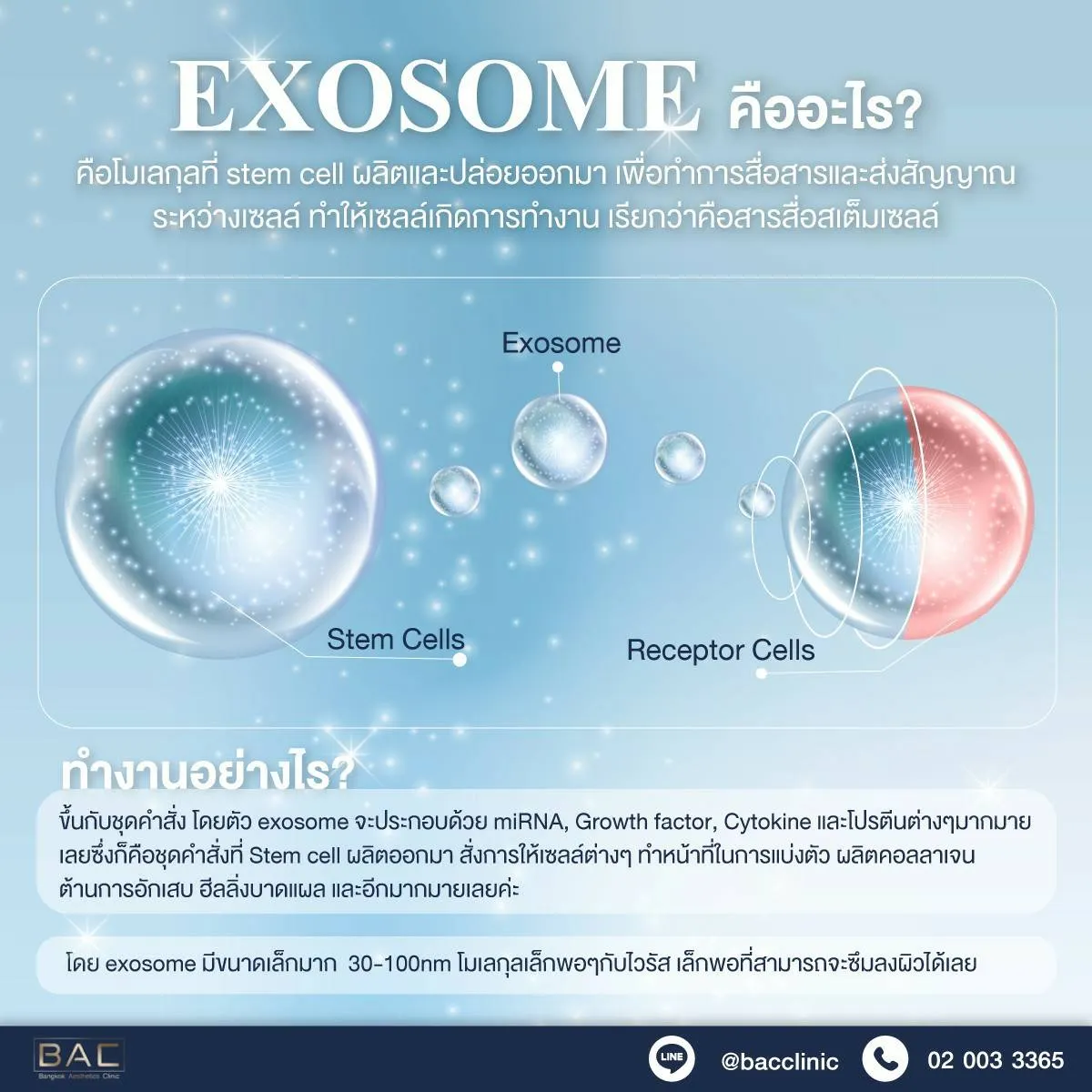
At BAC Clinic, we utilize cellular repair techniques by employing PRP-Regen Lab for clinical use in treating melasma. The starting price for PRP is 7,900 baht, while PRP-Regen Lab starts at 12,900 baht.
The clinical use of PRP-Regen Lab for treating melasma is supported by a study conducted by Çayırlı and colleagues in 2014.
In this study, the PRP-Regen Lab technique was utilized to treat melasma in patients aged 27, who had been experiencing melasma for approximately 5 years. Blood was extracted from the patients, approximately 8 milliliters, using test glass and centrifuged at a speed of 3,500 rpm for 8 minutes. The PRP-Regen Lab portion, located in the buffy coat area, was then separated. Using a 32 G needle, the PRP-Regen Lab was injected into the subdermal layer using a mesotherapy technique, targeting the papillary dermis at a depth of approximately 1.5 to 2.0 millimeters. Approximately 1.5 milliliters of PRP-Regen Lab was injected in total, divided into 3 sessions spaced 15 days apart. At the end of the study, epidermal melasma was reduced by approximately 80% without additional treatment, except for sunscreen application, as shown in Figure 2 (after).


The important components of blood flakes in α-granules contain over 30 biologically significant substances, including platelet-derived growth factor (PDGF), transforming growth factor (TGF)-β1, 2, epidermal growth factor, and mitogenic growth factors such as platelet-derived angiogenesis factor and fibrinogen. According to existing data, TGF-β1 is a growth factor associated with melasma development. Kim et al. studied the role of TGF-β1 in melasma development and found that TGF-β1 inhibits melanin production in a concentration-dependent manner, statistically significantly delaying extracellular signal-regulated kinase activation. In addition to the role of TGF-β1, the improvement in treatment outcomes may be related to skin thickening and strengthening, as PDGF plays a significant role in new blood vessel formation, collagen, and hyaluronic acid, components of the extracellular matrix. Hyaluronic acid has been shown to increase skin transparency and volume, contributing to skin rejuvenation. Therefore, the use of PRP-Regen Lab is an interesting option for individuals with melasma, as it not only reduces melanin production at the cellular level but also strengthens and rejuvenates the skin at the cellular level, reducing the likelihood of inflammation and thus reducing the recurrence of melasma.


Common abnormalities of pigmentation include:
- Freckles
- Sun spots or Solar lentigines

3. Hori’s Nevus
4. Melasma
5. Post-inflammatory hyperpigmentation (PIH)
“Pigmentation abnormalities encompass more than just freckles; they often involve a variety of issues.”

Dark spots, brown patches found on the skin have various names depending on the issue encountered, such as age spots, sunspots, uneven skin tone, freckles, and melasma. In one patient, multiple types of dark spots can be present, and although these problems may not pose a danger, they are generally unwelcome. These pigmentation issues often occur with age, especially after the age of 50, primarily due to sun exposure. Therefore, the best prevention is sunscreen application combined with the treatment of existing dark spots. Since dark spots have different names depending on the underlying factors, the choice of laser treatment varies. Thus, it is essential for physicians to diagnose and plan treatment to achieve the desired goals and reduce patient anxiety about their skin issues.
Factors that stimulate the formation of freckles and dark spots include hormonal changes, which can occur during pregnancy or when taking birth control pills. Freckle problems are particularly common during pregnancy, and abnormal pigmentation tends to fade after childbirth. For those experiencing freckles due to birth control pills, it’s advisable to consider alternative contraceptive methods to address this issue.
Regular application of sunscreen is essential. It’s recommended to choose a broad-spectrum sunscreen that protects against both UVA and UVB rays, with a minimum SPF of 15. Sunscreen should be applied at least 20 minutes before sun exposure and reapplied throughout the day. Nowadays, there are various sunscreen formats available, and using spray sunscreen can be convenient for reapplication during the day.
Medical treatment to reduce pigmentation abnormalities should be administered by a dermatologist. They will diagnose and prescribe appropriate treatments to suppress factors that stimulate freckle formation. Hydroquinone remains the gold standard for treating pigmentation abnormalities like freckles, sunspots, and dark spots, effectively reducing their intensity. However, caution must be exercised regarding potential side effects from long-term use. Combination therapy, such as combining hydroquinone with retinoids, is also an option. Additionally, other agents with lower efficacy levels, such as arbutin and vitamin C, can be used based on individual suitability.
Transamin, a medication discovered incidentally, is commonly used to treat freckles. It contains lysine, which inhibits melanin production and is involved in the suppression of the plasminogen activator process. However, its mechanism of action is not yet fully understood.
Chemical peels are another method of managing freckles, similar to laser treatment. Chemicals used for peeling, such as glycolic acid, lactic acid, and Jessner’s peel, affect the upper layers of the skin or the epidermis and dermis. However, it’s important to note that most pigmentation issues originate from deeper layers of the dermis or true skin. Therefore, using chemical peels that penetrate deeper layers may lead to side effects such as skin irritation, post-inflammatory hyperpigmentation (PIH), or dark spots, especially in Asian skin, which is more prone to such reactions.
When should freckles be treated with lasers?
Freckles or pigmentation abnormalities that resist treatment with freckle-reducing creams or oral medications for 6–8 weeks. Pigmentation abnormalities co-occurring with freckles, such as sunspots, melasma, or Hori’s nevus. When patients wish to rapidly eliminate freckles, such as before important events, and desire quick results, laser treatment should be considered as it provides faster results.
Testimonials


Treatment with lasers can be used alone or in combination with other treatments, and it’s not always necessary to use the same type of laser. This is because the factors contributing to the development of melasma vary from person to person. Additionally, the mechanism behind melasma formation isn’t entirely clear, including the level of abnormalities and skin layers affected, as well as the response to treatment. Other types of abnormal pigmentation may also differ, so treatment plans must be tailored by experienced medical professionals in collaboration with patients to prevent the recurrence of factors contributing to melasma and achieve rapid resolution, with reduced chances of recurrence.
Key Points about Laser Treatment for Melasma (LASER MELASMA TREATMENT FAQS):
Q: How can I tell if brown spots on my face are melasma or sun spots?
A: Melasma is often caused by hormonal changes, commonly occurring during pregnancy. Additionally, oral contraceptives can also induce melasma, especially in individuals sensitive to hormonal fluctuations. Changes in contraceptive brands can sometimes worsen melasma. Melasma induced by sun exposure, however, isn’t related to hormonal changes. Prolonged sun exposure, particularly as one ages, increases the risk of developing sun spots and melasma. Therefore, it’s important to avoid sun exposure by using sunscreen. Laser treatment for melasma caused by sun exposure is more challenging than treating sun-induced dark spots but can still be effective with the right approach and patient cooperation.
Q: Can red marks from acne be treated with lasers?
A: Red marks from acne result from inflammation during the acne phase and may be effectively treated with lasers since they’re not due to melanin abnormalities like melasma.
Q: If melasma occurs on the neck and spreads rapidly to areas like the arms and elbows, can treatment with creams containing hydroquinone and chemical peels not provide relief? Will laser treatment help?
A: BAC Clinic’s protocols for treating melasma and dark spots are more comprehensive and effective. Before treatment, the cause of melasma must be assessed to determine whether it’s stable or not. Different lasers with varying technologies should be used to evaluate the response of melasma to treatment, as different types of melasma respond differently. Therefore, proper evaluation by a specialized physician is crucial for selecting the most effective protocols tailored to the specific abnormalities in the skin layers.
Q: What are the current creams available for treating melasma and reducing dark spots?
A: The choice of cream for treating melasma and reducing dark spots depends on the severity of the condition. A combination of creams may be used, such as 2% hydroquinone along with azelaic acid, tranexamic acid, kojic acid, and, in severe cases, higher concentrations of hydroquinone. However, the selection of creams should be under medical supervision to prevent potential side effects.
In summary, laser treatment for melasma is a valuable option but should be part of a comprehensive treatment plan tailored to individual needs and under the supervision of experienced medical professionals.

References
Ephelides (freckles) — Dermatology Advisor. (n.d.). Retrieved January 11, 2022, from https://www.dermatologyadvisor.com/home/decision-support-in-medicine/dermatology/ephelides-freckles/
Lee, Y. J., Shin, H. J., Noh, T.-K., Choi, K.-H., & Chang, S.-E. (2017). Treatment of melasma and post-inflammatory hyperpigmentation by a picosecond 755-nm alexandrite laser in Asian patients. Annals of Dermatology, 29(6), 779–781.
Solar lentigines — dermoscopedia. (n.d.). Retrieved January 11, 2022, from https://dermoscopedia.org/Solar_lentigines
Tian, B. W. C. A. (2015). Novel treatment of Hori’s nevus: A combination of fractional nonablative 2,940-nm Er: YAG and low-fluence 1,064-nm Q-switched Nd: YAG laser. Journal of Cutaneous and Aesthetic Surgery, 8(4), 227.